Home

Emmek okuyucu Postane koedam kappa işaret fişeği hasta Doğurmak

Visual assessment of the medial temporal lobe atrophy was performed on... |  Download Scientific Diagram
Visual assessment of the medial temporal lobe atrophy was performed on... | Download Scientific Diagram

AVRA: Automatic visual ratings of atrophy from MRI images using recurrent  convolutional neural networks
AVRA: Automatic visual ratings of atrophy from MRI images using recurrent convolutional neural networks
Analysis of regional atrophy on brain imaging compared with cognitive  function in the elderly and in patients with dementia –
Analysis of regional atrophy on brain imaging compared with cognitive function in the elderly and in patients with dementia –

AVRA: Automatic visual ratings of atrophy from MRI images using recurrent  convolutional neural networks
AVRA: Automatic visual ratings of atrophy from MRI images using recurrent convolutional neural networks

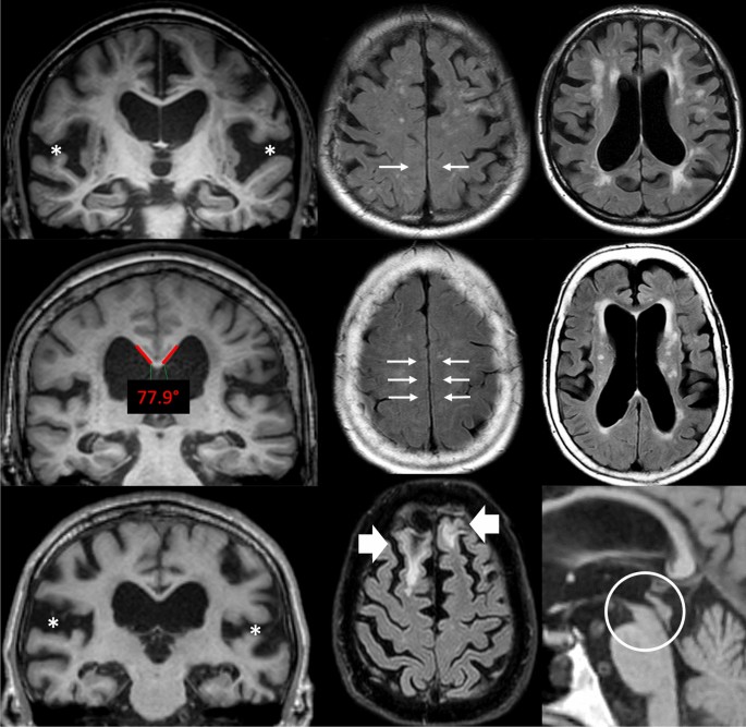
Imaging features associated with idiopathic normal pressure hydrocephalus  have high specificity even when comparing with vascular dementia and  atypical parkinsonism | Fluids and Barriers of the CNS | Full Text
Imaging features associated with idiopathic normal pressure hydrocephalus have high specificity even when comparing with vascular dementia and atypical parkinsonism | Fluids and Barriers of the CNS | Full Text

AVRA: Automatic visual ratings of atrophy from MRI images using recurrent  convolutional neural networks - ScienceDirect
AVRA: Automatic visual ratings of atrophy from MRI images using recurrent convolutional neural networks - ScienceDirect

Tips for teachers of evidence-based medicine: 3. Understanding and  calculating kappa
Tips for teachers of evidence-based medicine: 3. Understanding and calculating kappa

AVRA: Automatic visual ratings of atrophy from MRI images using recurrent  convolutional neural networks
AVRA: Automatic visual ratings of atrophy from MRI images using recurrent convolutional neural networks

PDF) Visual assessment of posterior atrophy development of a MRI rating  scale
PDF) Visual assessment of posterior atrophy development of a MRI rating scale

Julie Koedam, RHIT - Coding Quality Coordinator/Auditor - BJC HealthCare |  LinkedIn
Julie Koedam, RHIT - Coding Quality Coordinator/Auditor - BJC HealthCare | LinkedIn

Parieto-occipital sulcus widening differentiates posterior cortical atrophy  from typical Alzheimer disease - ScienceDirect
Parieto-occipital sulcus widening differentiates posterior cortical atrophy from typical Alzheimer disease - ScienceDirect

AVRA: Automatic visual ratings of atrophy from MRI images using recurrent  convolutional neural networks - ScienceDirect
AVRA: Automatic visual ratings of atrophy from MRI images using recurrent convolutional neural networks - ScienceDirect

PDF) Parieto-occipital sulcus widening differentiates posterior cortical  atrophy from typical Alzheimer disease
PDF) Parieto-occipital sulcus widening differentiates posterior cortical atrophy from typical Alzheimer disease

Voxel-wise deviations from healthy aging for the detection of  region-specific atrophy - ScienceDirect
Voxel-wise deviations from healthy aging for the detection of region-specific atrophy - ScienceDirect

Posterior Atrophy and Medial Temporal Atrophy Scores Are Associated with  Different Symptoms in Patients with Alzheimer's Disease and Mild Cognitive  Impairment | PLOS ONE
Posterior Atrophy and Medial Temporal Atrophy Scores Are Associated with Different Symptoms in Patients with Alzheimer's Disease and Mild Cognitive Impairment | PLOS ONE

PDF) Top 10 Helpful Measurements and Scoring System for Neurodegenerative  Brain MRI Interpretation
PDF) Top 10 Helpful Measurements and Scoring System for Neurodegenerative Brain MRI Interpretation

Structural imaging findings on non-enhanced computed tomography are  severely underreported in the primary care diagnostic work-up of subjective  cognitive decline | SpringerLink
Structural imaging findings on non-enhanced computed tomography are severely underreported in the primary care diagnostic work-up of subjective cognitive decline | SpringerLink

PDF) Evaluation of MR Visual Rating Scales in Major Forms of Dementia

PDF) Evaluation of MR Visual Rating Scales in Major Forms of Dementia

Automated quantitative MRI volumetry reports support diagnostic  interpretation in dementia: a multi-rater, clinical accuracy study |  SpringerLink
Automated quantitative MRI volumetry reports support diagnostic interpretation in dementia: a multi-rater, clinical accuracy study | SpringerLink

Tips for learners of evidence-based medicine: 3. Measures of observer  variability (kappa statistic). - Abstract - Europe PMC
Tips for learners of evidence-based medicine: 3. Measures of observer variability (kappa statistic). - Abstract - Europe PMC

Visual assessment of posterior atrophy development of a MRI rating scale |  SpringerLink
Visual assessment of posterior atrophy development of a MRI rating scale | SpringerLink

Deep learning for chest radiograph diagnosis: A retrospective comparison of  the CheXNeXt algorithm to practicing radiologists | PLOS Medicine
Deep learning for chest radiograph diagnosis: A retrospective comparison of the CheXNeXt algorithm to practicing radiologists | PLOS Medicine
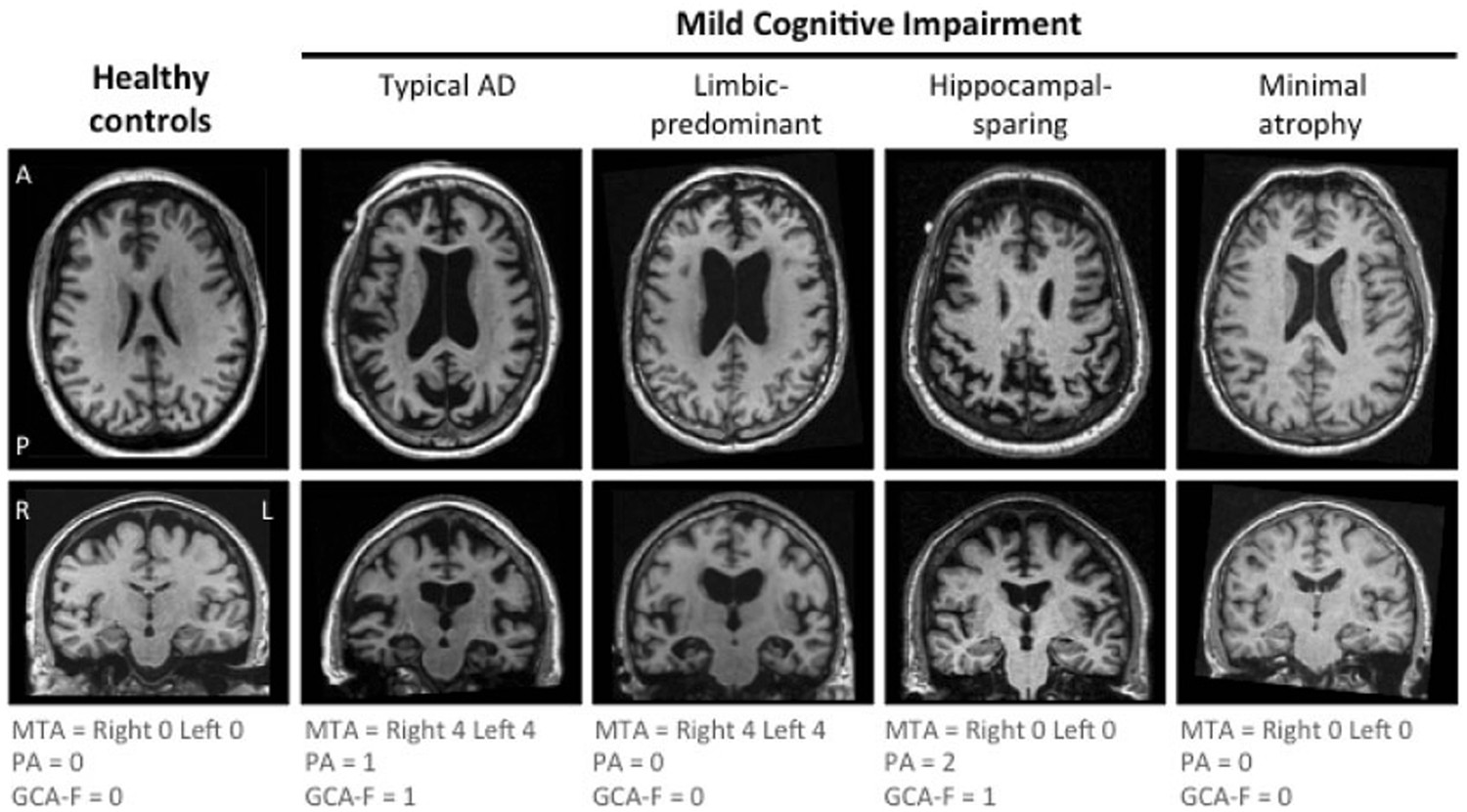
The A/T/N biomarker scheme and patterns of brain atrophy assessed in mild  cognitive impairment | Scientific Reports
The A/T/N biomarker scheme and patterns of brain atrophy assessed in mild cognitive impairment | Scientific Reports

Posterior cerebral atrophy in the absence of medial temporal lobe atrophy  in pathologically-confirmed Alzheimer's disease - ScienceDirect
Posterior cerebral atrophy in the absence of medial temporal lobe atrophy in pathologically-confirmed Alzheimer's disease - ScienceDirect